For decades, structural biologists have relied on specialized file formats to store and exchange the detailed three-dimensional coordinates of macromolecules. Among these, the most widely recognized are the classic PDB (Protein Data Bank) format and its modern successor, the CIF (Crystallographic Information File).
The PDB format was revolutionary for its time, providing a standardized way to represent molecular structures and share them across the scientific community. However, as structural biology advanced and the complexity of solved models increased, the limitations of the PDB format became increasingly evident. To address these challenges, the CIF format was introduced as a more versatile, extensible, and data-rich alternative.
In this article, we explore the origins, structure, and fundamental differences between the PDB and CIF formats, explaining why the global structural biology community is progressively shifting toward CIF. Understanding these distinctions will help you determine which format best suits your research or computational needs.
A Deep Dive into the PDB Format
Introduced in the 1970s, the PDB (Protein Data Bank) format served as the foundational standard for storing 3D coordinate data of biological macromolecules such as proteins and nucleic acids. Its straightforward and rigid design made it widely accessible during the early development of computational biology.

The format follows a fixed-width column structure, inspired by the 80-column punch cards used in early computing. Each data point—atom number, atom name, residue name, chain identifier, and X, Y, Z coordinates—must appear in exact column positions. While this rigidity ensures consistency, it also imposes significant constraints as molecular data becomes more complex.
Key Record Types:
- HEADER: Contains general information about the molecular entry.
- ATOM: Stores coordinate information for standard atoms within proteins or nucleic acids.
- HETATM: Describes coordinates for heteroatoms (non-polymer components) such as ligands, ions, or water molecules.
- TER: Marks the termination of a polymer chain.
PDB File Example
Here is a small snippet showing a few atoms of the amino acid Alanine. Notice the strict alignment of data into columns:
ATOM      1  N   ALA A   1      -1.496  -0.347   0.316  1.00 13.79           N
ATOM      2  CA  ALA A   1      -0.129  -0.016  -0.081  1.00 12.82           C
ATOM      3  C   ALA A   1       0.871  -1.123   0.091  1.00 12.98           C
ATOM      4  O   ALA A   1       0.569  -2.223  -0.329  1.00 14.38           O
ATOM      5  CB  ALA A   1       0.149   1.321   0.568  1.00 12.39           C
To visualize such coordinates in 3D, researchers often use online PDB viewers, which allow users to upload a PDB file and interactively explore its molecular structure.
Key Limitations of the PDB Format
Despite its historic significance, the PDB format suffers from several technical limitations:
- Atom Count Restriction: The format supports a maximum of 99,999 atoms, making it unsuitable for large biological assemblies such as ribosomes or viral capsids.
- Chain Identifier Limit: Each chain can be labeled with only one character (A–Z, 0–9), restricting the number of unique chains per file.
- Poor Extensibility: Incorporating additional metadata or novel data types is extremely challenging due to its rigid, column-based structure.

These limitations prompted the development of more flexible and information-rich alternatives—most notably, the CIF format. Despite its limitations, the PDB format remains widely used. If you need to convert a PDB file to another format for a specific analysis pipeline, you can use a dedicated online tool like our PDB File Converter.
Choose the PDB Format When:
- You work with legacy software that does not support CIF.
- You are visualizing small, simple structures where limitations are not critical.
- You are teaching basic atomic coordinate concepts in an educational setting.
Defining the CIF Format
To address the limitations of the PDB format, the International Union of Crystallography (IUCr) developed the CIF (Crystallographic Information File) format. For macromolecules such as proteins and nucleic acids, an extended version known as mmCIF (macromolecular CIF) is used. This format is now the official and mandatory standard for all new structure submissions to the Worldwide Protein Data Bank (wwPDB).
For simplicity, we refer to both CIF and mmCIF collectively as CIF throughout this guide. (For a detailed comparison, see CIF vs. mmCIF: Understanding the Difference.)
Unlike PDB, CIF uses a flexible tag-value (or key-value) structure. Each piece of data is preceded by a descriptive tag (e.g., _atom_site.Cartn_x for the X coordinate). This self-describing nature makes the format far easier for both humans and computers to parse. For tabular data, such as a list of atoms, CIF uses a loop_ structure where tags are defined once as column headers, followed by rows of data.

Example of a CIF File
Below is the same Alanine structure represented in CIF format. Notice how the information is structured using tag-value pairs and the loop_ keyword:
loop_
_atom_site.group_PDB
_atom_site.id
_atom_site.type_symbol
_atom_site.label_atom_id
_atom_site.label_comp_id
_atom_site.auth_asym_id
_atom_site.auth_seq_id
_atom_site.Cartn_x
_atom_site.Cartn_y
_atom_site.Cartn_z
_atom_site.occupancy
_atom_site.B_iso_or_equiv
ATOM   1 N N  ALA A 1 -1.496 -0.347  0.316 1.00 13.79
ATOM   2 C CA ALA A 1 -0.129 -0.016 -0.081 1.00 12.82
ATOM   3 C C  ALA A 1  0.871 -1.123  0.091 1.00 12.98
ATOM   4 O O  ALA A 1  0.569 -2.223 -0.329 1.00 14.38
ATOM   5 C CB ALA A 1  0.149  1.321  0.568 1.00 12.39
#
To visualize CIF data in 3D, researchers often use interactive CIF viewers.
Key Advantages of the CIF Format
- No Size Limit: CIF can handle any number of atoms, chains, or data categories without restriction.
- Highly Flexible: The tag-value structure makes it easy to incorporate rich metadata, including experimental conditions, refinement parameters, and validation reports.
- Official Standard: As the wwPDB’s official format, CIF is universally supported by modern molecular visualization and analysis software.

Choose the CIF Format When:
- You are analyzing or depositing modern, complex structures such as ribosomes or viruses.
- You are submitting new data to the Protein Data Bank.
- You require comprehensive experimental metadata for reproducibility or analysis.
- You are writing computational scripts—CIF’s format is more reliable and easier to parse.
- You are using AI-based prediction tools like AlphaFold3, which export their results in CIF format.
As the modern standard, you’ll frequently work with CIF files. For situations where you need to ensure compatibility with older software or different tools, a flexible converter can be invaluable. You can convert CIF files to the legacy PDB format or other types using a tool like the CIF File Converter.
PDB vs. CIF: A Side-by-Side Comparison
| Feature | PDB Format | CIF Format |
|---|---|---|
| Structure | Fixed-Width Columns | Tag-Value Pairs |
| Size Limit | Yes (99,999 atoms) | No |
| Flexibility | Low | High |
| Metadata | Basic | Rich and Detailed |
| Current Standard | Legacy | Official Standard |
How to Convert CIF to PDB Format?
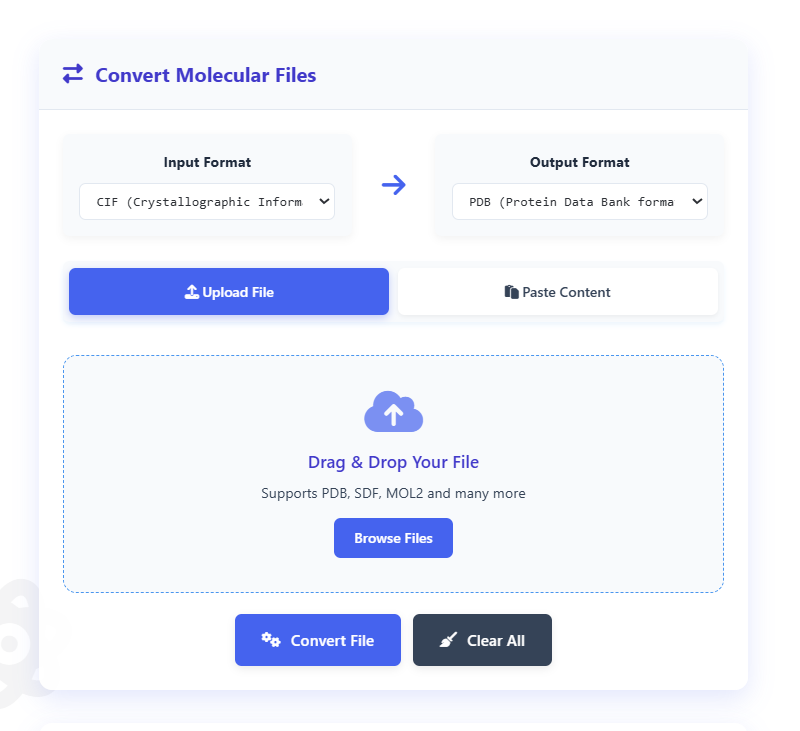
While CIF is the modern standard, you may occasionally need to convert a CIF file back to the legacy PDB format, especially for older software. It’s crucial to remember that this conversion is only possible if the structure fits within the PDB format’s limitations (e.g., fewer than 99,999 atoms), and some metadata will likely be lost. This conversion can often be done with online tools, such as the CIF to PDB Converter.
Conclusion
The PDB format was a foundational tool that helped launch the field of structural biology, and its importance cannot be overstated. While the scientific community has adopted the more scalable and flexible CIF format as the official standard for archiving new and complex structures, the PDB format’s era is far from over. It remains deeply integrated into countless software programs and established analysis pipelines. Consequently, for reasons of compatibility and simplicity with smaller structures, the PDB format continues to be a practical and essential tool for many researchers. Ultimately, a modern structural biologist must be proficient with both formats to navigate the field effectively.
References
- Berman, H. M., Westbrook, J., Feng, Z., Gilliland, G., Bhat, T. N., Weissig, H., … & Bourne, P. E. (2000). The Protein Data Bank. Nucleic Acids Research, 28(1), 235-242.
- Hall, S. R., Allen, F. H., & Brown, I. D. (1991). The Crystallographic Information File (CIF): a new standard archive file for crystallography. Acta Crystallographica Section A: Foundations of Crystallography, 47(6), 655-685.
- Bourne, P. E., Westbrook, J., Berman, H. M., Fitzgerald, P. M. D., & IUCr. (1997). The Macromolecular Crystallographic Information File (mmCIF). Methods in Enzymology, 277, 571-590.
- Westbrook, J. D., & Bourne, P. E. (2000). The impact of the mmCIF format on the Protein Data Bank. Bioinformatics, 16(2), 159-168.