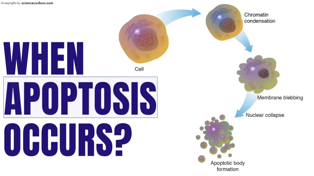
The term “apoptosis” was first introduced by researcher Kerr in 1972 to describe physiological cell death based on morphological changes and to distinguish it from necrosis. Apoptosis is a Greek word meaning “falling off” or “dropping off.” The terms “apoptosis” and “programmed cell death” are often used interchangeably. In contrast, others have referred to apoptosis as the most significant form of programmed cell death. In this article, we will examine the importance of apoptosis in our body.
What Happens During Apoptosis?

Apoptosis is a form of programmed cell death that occurs in multicellular organisms and some eukaryotic microorganisms. Apoptosis is a series of biochemical events that lead to chromatin condensation and fragmentation, ultimately resulting in cell death (cell suicide). Apoptosis occurs when DNA damage is irreparable, the cell is infected with a virus, or the cell is under severe stress.
At the molecular level, apoptosis can be triggered in two main ways: through signals on the cell’s surface (extrinsic pathway) or internal stress signals from mitochondria (intrinsic pathway), controlled by proteins like Bcl-2 and enzymes called caspases. When apoptosis works as it should, it keeps tissues balanced and even shapes our bodies during development—for example, helping separate fingers and toes in the womb. But when it goes wrong, problems arise: too little apoptosis can let cancer cells survive, while too much can drive conditions like Alzheimer’s or heart disease.

Apoptosis occurs when:
- Cell pollution by pathogenic agents such as viruses, bacteria, or fungi
- When A cell has become badly stressed or irreparably damaged.
- Performing mutations in genetic material and the inability to repair and correct the mutation performed, the cell is senescent.
- Another factor in cell apoptosis is the accumulation of damaged and dysfunctional proteins in the rough endoplasmic reticulum.
- Shortening the length of the telomere region of genetic material can cause cell apoptosis.
- Completion of growth and development: Some cells must be removed during growth and development, which can be observed in the removal of tissue between the fingers during embryonic development.
- A cell’s existence is no longer helpful to the organism.
- Cells have been in the body long enough that they’re worn out, so they need to be replaced by nice, new, young cells.
- Brain cells undergo apoptosis before and following birth to eliminate excess brain cells and streamline nerve impulses.
- Triggered by mild cellular injury and by various factors, internal or external to the cell
Apoptosis in Body Homeostasis
In adult multicellular animals, the number of body cells usually remains constant. This constancy is due to the coordination of mitosis and programmed cell death. Cells create new cells through division. On the other hand, old, damaged, or excess cells are removed through apoptosis. For further explanation, let’s consider an example. B lymphocytes proliferate and increase after encountering an antigen.
These cells often succeed in removing foreign antigens by producing antibodies. However, after the foreign antigen is removed, the body does not need many B lymphocytes produced in response to it. So, what will happen to the stimulated B lymphocytes? The answer is very simple. These cells are removed by apoptosis after the antigen is removed. Of course, some B lymphocytes remain memory cells to defend the body against the same antigen in future encounters.
Apoptosis in Development
Programmed cell death (apoptosis) is essential for shaping tissues and organs during the growth of both animals and plants. It’s not just about removing “unwanted” cells—it’s a finely tuned process that ensures proper body structure. In embryological development, apoptosis acts like a sculptor. Research on chicken embryos shows that certain cells are selectively removed through apoptosis, a pattern in many other organisms. This process is tightly regulated, with different tissues relying on specific signals to eliminate extra or misplaced cells. For example, in birds, bone morphogenetic proteins (BMPs) trigger apoptosis in the cells between developing digits, so fingers and toes separate instead of staying webbed. In fruit flies (Drosophila), steroid hormones initiate apoptosis to clear away excess, damaged, or old cells during development.
FAQ
What is apoptosis in simple terms?
Apoptosis is a natural way for the body to eliminate old, damaged, or unnecessary cells. Think of it as a “cell retirement program” that keeps tissues healthy.
How is apoptosis different from necrosis?
Apoptosis is a clean, controlled process where cells quietly break down and are removed without causing inflammation. Necrosis, on the other hand, happens when cells die due to injury or stress, often leading to swelling, leakage, and inflammation.
Why is apoptosis important during development?
Apoptosis shapes the body during growth. For example, it helps separate fingers and toes in an embryo by removing the cells in between. Without apoptosis, proper development wouldn’t be possible.
Can problems with apoptosis cause diseases?
Yes. Too little apoptosis allows damaged or mutated cells to survive, which can lead to cancer. Too much apoptosis causes the loss of healthy cells, which is linked to diseases like Alzheimer’s, Parkinson’s, and heart failure.
How do scientists study apoptosis?
Researchers use lab techniques such as flow cytometry, staining methods (like Annexin V or TUNEL assay), and imaging to track the stages of apoptosis and understand how it works.
Can apoptosis be targeted for treatment?
Absolutely. Many cancer therapies are designed to trigger apoptosis in tumor cells, while treatments for neurodegenerative diseases aim to slow down excessive apoptosis to protect healthy cells.
This article was medically reviewed for accuracy by Dr. Zeinab Morshedi Yekta. The content is based on scientific evidence and is intended to be educational. It does not replace professional medical advice.
Conclusion
In conclusion, apoptosis, or programmed cell death, is a vital biological process that ensures multicellular organisms’ health, balance, and proper development. By carefully eliminating damaged, unnecessary, or excess cells, apoptosis maintains tissue homeostasis, shapes our bodies during embryonic development, and protects against diseases like cancer when functioning correctly. However, its dysregulation can contribute to severe conditions, highlighting its delicate yet critical role. From sculpting fingers in the womb to clearing out worn-out cells in adulthood, apoptosis is a remarkable example of nature’s precision, quietly orchestrating life’s balance behind the scenes.
Reference
- Singh, P., & Lim, B. (2022). Targeting apoptosis in cancer. Current oncology reports, 24(3), 273-284.
- Mustafa, M., Ahmad, R., Tantry, I. Q., Ahmad, W., Siddiqui, S., Alam, M., … & Islam, S. (2024). Apoptosis: a comprehensive overview of signaling pathways, morphological changes, and physiological significance and therapeutic implications. Cells, 13(22), 1838.
- Lossi, L. (2022). The concept of intrinsic versus extrinsic apoptosis. Biochemical Journal, 479(3), 357-384.
- Biorender.com