Echinococcus multilocularis, also known as the fox tapeworm, is a parasitic worm that causes alveolar echinococcosis (AE), a serious and potentially fatal disease in humans.
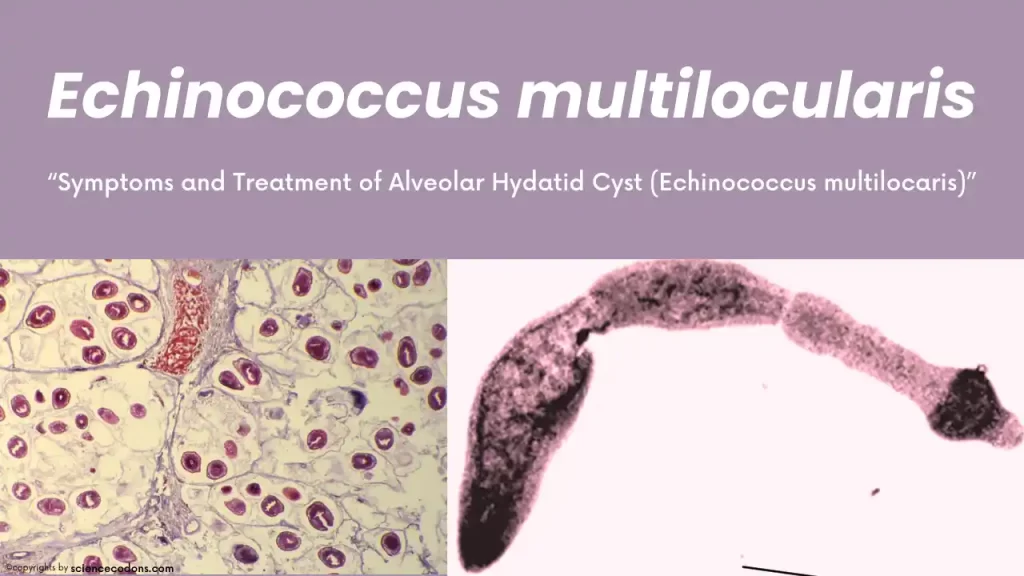
Echinococcus multilocularis is a type of cestode that belongs to the Cyclophyllidea order and the Taeniidae family. This tapeworm is responsible for causing Alveolar hydatid cysts. While Echinococcus granulosus causes hydatid cysts, the hydatid cysts created by Echinococcus multilocularis are alveolar, which distinguishes these two types of worms.
What is E. multilocularis? [Morphology & Characteristics]
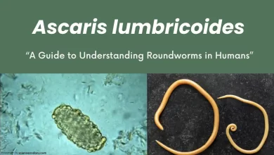
Echinococcus multilocularis is known to cause the most dangerous worm infection in humans. This small worm measures between 1.2 to 3.7 millimeters and has 3-5 segments. Its eggs are similar to other tapeworms, but these eggs can withstand cold temperatures up to minus 26 degrees Celsius for 60 days. This means that even freezing cannot eliminate this worm.
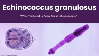
The Alveolar multi-locular hydatid cyst, which is the larval stage of E. multilocularis, is found in the northern hemisphere regions such as Canada, Russia, China, Turkey, and northeastern Iran (Moghan Plain). The scolex of the worm is equipped with 4 suckers, a rostellum, and 2 rows of hooks. It has a short neck, an immature segment, a mature segment, and a nearly large gravid segment. Overall, it is similar to granulosus, but they differ in terms of mature and gravid segments, the number of testes and their location, and the location of the genital pore. This worm measures between 3.5 to 4 millimeters.
Lifecycle and Transmission
The definitive host of Echinococcus granulosus is the red fox and its intermediate hosts are mice and rodents. Humans can also be accidental intermediate hosts. The adult worm lives in the intestine of the definitive host (fox). Segments containing eggs are excreted and consumed by the intermediate host. When the intermediate host eats these eggs with feces, the six-hooked embryo comes out of the egg and migrates to the visceral organs through the bloodstream.
The liver is the organ that is most involved in the first stage. A cyst is formed in this organ and proliferates to such an extent that it metastasizes to other organs and encompasses the entire abdominal area, causing the death or illness of the rodent. If the definitive host hunts this rodent, the protoscoleces of Echinococcus multilocularis are usually released from the cyst and their heads attach to the intestinal wall through suckers and hooks. The worm matures and begins to excrete gravid segments. Humans become infected by eating eggs that the fox excretes. The six-hooked embryo is released in the intestine and enters the internal organs of the abdomen, especially the liver, through the bloodstream, where it forms a cyst that can show its symptoms.
| Life Cycle Stage | Description |
|---|---|
| Adult | Resides in the small intestine of the definitive host. Gravid proglottids release eggs that are passed in the feces, and these eggs are immediately infectious. |
| Eggs | After ingestion by a suitable intermediate host, eggs hatch in the small intestine and release six-hooked oncospheres. These oncospheres penetrate the intestinal wall and migrate through the circulatory system into various organs, primarily the liver. |
| Hydatid Cyst | In the liver or lungs, the oncosphere develops into a thick-walled hydatid cyst that gradually enlarges. The cyst produces protoscolices and daughter cysts within its interior. |
| Transmission to Definitive Host | The definitive host becomes infected by ingesting the cyst-containing organs of the infected intermediate host. After ingestion, the protoscolices attach to the intestinal mucosa and develop into adult stages within 32 to 80 days. |
| Humans as Aberrant Intermediate Hosts | Humans can become infected by ingesting eggs. Oncospheres are released in the intestine, and hydatid cysts develop in various organs. If cysts rupture, liberated protoscolices may create secondary cysts in other sites within the body (secondary echinococcosis). |
In Echinococcus multilocularis, eggs can create multiple cysts (alveolar and vesicular); while a hydatid cyst is a vesicle that may contain several daughter cysts. Also, these two differ in their definitive and intermediate hosts.
Symptoms of Echinococcus multilocularis
The first organ that Echinococcus multilocularis affects is the liver. Statistics indicate that in 28% of cases, the right lobe of the liver is involved, in 9% of cases, the left lobe, and in 63% of cases, both lobes of the liver are affected. The most common symptoms of Alveolar hydatid cysts in humans are intermittent and constant abdominal pain, and obstructive jaundice due to pressure on the bile ducts causing their obstruction.
| Affected Organ | Symptoms |
|---|---|
| Liver | Abdominal pain, yellowing of the skin, and whites of the eyes (jaundice) if cysts form in the liver. |
| Lungs | Chest pain and coughing up blood or the contents of cysts if cysts form in the lungs. |
The organs that are most often metastasized are the lungs and brain. These do not have a cystic appearance (their touch and texture are hard). They mimic malignant tumors and appear similar to tumors. They are white-gray in color. Usually, there is one, and later the number increases, metastasizes, and in the liver, it appears exactly like malignant diseases and cancer.
Diagnosis and treatment of E. multilocularis
In the initial diagnosis of Echinococcus multilocularis, it often appears as a cancer of the digestive system and liver. In radiography, eosinophilia, lymphocytopenia, and calcified rings are seen. We can use serological techniques like ELISA to search for the worm’s antibodies in the patient’s blood and imaging techniques such as CT scan and MRI, similar to what is used for granulosus (hydatid cyst).
The main treatment for Echinococcus multilocularis is radical surgery (multi-stage) (the cyst must be surgically removed and removed in several stages because the number of cysts is not one to be cured with one surgery). In drug treatment, it does not respond well to praziquantel, but in some cases, it responds well to albendazole (mebendazole). The albendazole dose is recommended as 40 milligrams per kilogram for 6 months.
Prevention and Control
Prevention strategies focus on reducing the risk of human exposure to the eggs of E. multilocularis. This includes:
- Regular deworming of domestic dogs.
- Safe handling and disposal of animal carcasses.
- Washing hands after contact with dogs or potential exposure to contaminated soil.
- Ensuring that food and water are free from contamination.
Also, it is recommended to eliminate rodents and avoid them and thoroughly wash fruits and vegetables that are not contaminated with worm eggs. This worm is very dangerous, and existing treatments are not completely effective, so the best way to deal with this worm is prevention.
Conclusion
Echinococcus multilocularis poses a significant health threat, particularly in endemic areas. Awareness and preventive measures are key to controlling the spread of this parasite and protecting public health. It is essential for individuals in at-risk areas to be aware of the risks and to take steps to prevent infection.
Reference
- CDC
- DOI: 10.5772/intechopen.68565
- Mihmanli M, Idiz UO, Kaya C, Demir U, Bostanci O, Omeroglu S, Bozkurt E. Current status of diagnosis and treatment of hepatic echinococcosis. World Journal of Hepatology. 2016;8:1169-1181. DOI: 10.4254/wjh.v8.i28.1169
- Hu D, Song X, Xie Y, Zhong X, Wang N, Zheng Y, Gu X, Wang T, Peng X, Yang G. Molecular insights into a tetraspanin in the hydatid tapeworm Echinococcus granulosus. Parasites & Vectors. 2015;8:311. DOI: 10.1186/s13071‐015‐0926‐y
- Kouguchi H, Matsumoto J, Nakao R, Yamano K, Oku Y, Yagi K. Characterization of a surface glycoprotein from Echinococcus multilocularis and its mucosal vaccine potential in dogs. PLoS One. 2013;8:e69821. DOI: 10.1371/journal.pone.0069821
- uan M, Luo Y, Xin Q, Gao H, Zhang G, Jing T. Efficacy of osthole for Echinococcus granulosus in vitro and Echinococcus multilocularis in vivo. Veterinary Parasitology. 2016;226:38-43. DOI: 10.1016/j.vetpar.2016.05.016
- Journal of Helminthology
- doi.org/10.1186/s13071-016-1746-4