Clonorchis sinensis, also known as the Chinese liver fluke, is a type of liver fluke. This parasite affects the liver and bile ducts, potentially causing gallbladder inflammation and cholangitis. Interestingly, it can mimic the symptoms of gallstones, as both this worm and gallstones can cause obstructions. In some cases, infection with this worm can even lead to bile duct cancer or cholangiosarcoma.
What is Clonorchis sinensis? [Morphology & Characteristics]
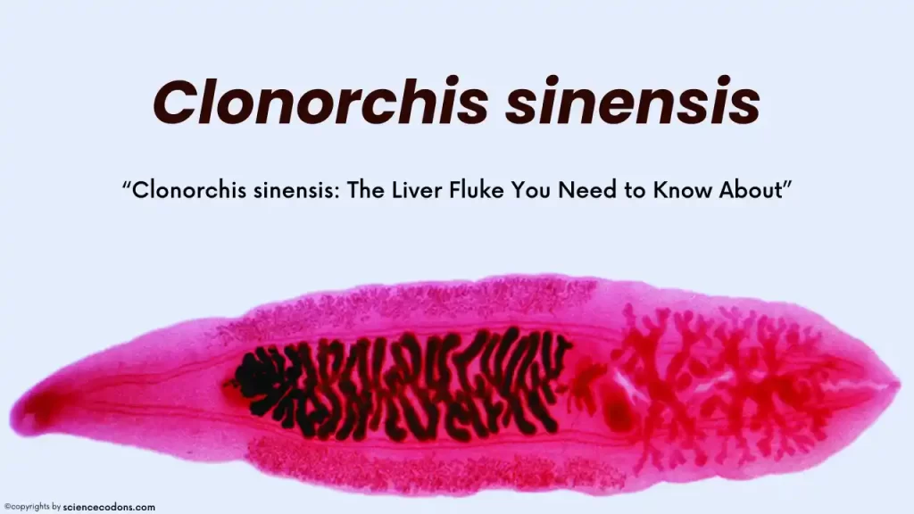
Clonorchis sinensis is commonly referred to as the Chinese liver worm. Its primary geographical distribution is in the Far East, particularly in China. The worm has a lancet-like appearance, with a smooth, golden-brown body covered in cuticle. It measures between 1 to 2.5 centimeters in length and 3-5 millimeters in width. The worm’s oral sucker is larger than its ventral sucker.
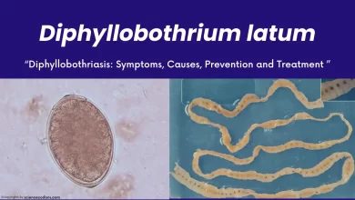
Clonorchis sinensis typically has a blind intestine (cecum) closed at the end and without branches. Its testes and ovaries are also branched. The reproductive system is arranged in an OTT (ovary up and testes down) format. The eggs measure about 20 to 30 microns and have a clear operculum (valve). They also have a knob or appendage seen at the end of the pole opposite the valve. The eggs are mature, and these features can be used to diagnose this parasite in collected samples.
Geographic Distribution
Endemic to regions such as Korea, China, Taiwan, and northern Vietnam, Clonorchis sinensis is Asia’s most prevalent human fluke and the third-most globally. The majority of infections are reported in China.
The life cycle of Clonorchis sinensis
This worm can live in the human body for 20-25 years. It resides in the liver and bile ducts. The eggs enter the intestine along with the bile and are excreted through the feces. These eggs are mature. When the first intermediate host, the snail, eats the eggs, the miracidium comes out of the egg and goes through the sporocyst, rediae, and cercaria stages in the snail’s soft tissues.
The cercaria leaves the snail’s body, swims with its tail, and enters the second host, fish. The cercariae of Clonorchis sinensis turns into the next larval stage, metacercaria, in the fish’s muscles. If the infected fish is consumed raw or half-cooked by humans, after the fish muscles are digested in the digestive system, the metacercaria opens, and the young worm comes out of the metacercaria and migrates to the bile ducts of the liver. Dogs, cats, pigs, and mice can also be the final hosts.
| Stage | Description |
|---|---|
| Adult | Resides in the bile ducts of humans or other mammals |
| Eggs | Released into the environment via feces |
| Miracidia | Hatch from eggs and infect freshwater snails |
| Sporocysts | Develop within snails |
| Cercariae | Emerge from snails and infect freshwater fish |
| Metacercariae | Encyst in the flesh of fish |
| Ingestion | Humans or other mammals eat infected fish |
Clinical Symptoms in C. Sinensis Infection
Mild infections caused by Clonorchis sinensis typically do not present any symptoms. However, we may observe adenomatous changes in the bile ducts, including thickening, stretching, and fibrosis.
Severe infections can manifest more severe symptoms such as jaundice, hepatomegaly (enlarged liver), abdominal pain, diarrhea, loss of appetite, inflammation of the gallbladder and bile ducts (cholangitis), and an increased risk of bile duct cancer, which is a significant concern and is referred to as cholangiocarcinoma.
In chronic cases, patients may experience liver-related symptoms associated with a large number of worms, often due to recurrent infections.
| Stage | Symptoms |
|---|---|
| Acute | Fever, abdominal pain, hepatomegaly, eosinophilia |
| Chronic | Cholangitis, cholecystitis, liver abscess, pancreatitis |
| Advanced | Obstructive jaundice, cholangiocarcinoma |
Diagnosis and Treatment C. sinensis
Clonorchis sinensis is diagnosed through stool testing and observing the eggs in the stool. A stool sample is taken, then a swab is taken from it after dilution and placed on a slide. The sample is then examined under a light microscope. Several slides may be prepared from each sample, and all will be examined. It’s important to note that the eggs of Clonorchis cannot be distinguished from the eggs of Heterophyes and Metagonimus due to their high similarity. The primary treatment for this and other flatworm infections is using praziquantel.
Conclusion
Clonorchis sinensis is a significant food-borne parasite linked to serious hepatobiliary diseases, including cholangiocarcinoma and hepatocellular carcinoma (HCC). Recent studies have shown that infection with C. sinensis can worsen the prognosis for patients with HCC by promoting cancer stem cell-like characteristics, leading to accelerated malignant progression. This highlights the importance of addressing C. sinensis infections as a public health priority, particularly in endemic regions, to prevent associated complications and improve patient outcomes. Efforts toward better diagnosis, treatment, and prevention strategies are essential to combat the impact of this parasite on global health.
Reference
- Qian MB, Chen YD, Yan F. Time to tackle clonorchiasis in China. Chin J Parasitol Parasit Dis. 2010;28(2):113-8.
- Hong ST, Fang Y. Clonorchis sinensis and clonorchiasis, an update. Parasitol Int. 2012 Mar;61(1):17-24. [PubMed: 21741496]
- ncbi.nlm.nih.gov/books/NBK532892/
- WHO