Echinococcus granulosus is a tapeworm from the Taeniidae family. This worm was first reported by Hartman in 1690. It is known to cause hydatid cysts, which are significantly prevalent and can be fatal.
Echinococcus granulosus, commonly known as the hydatid worm or dog tapeworm, is a parasitic organism responsible for causing cystic echinococcosis in humans and animals
Morphology of Echinococcus granulosus
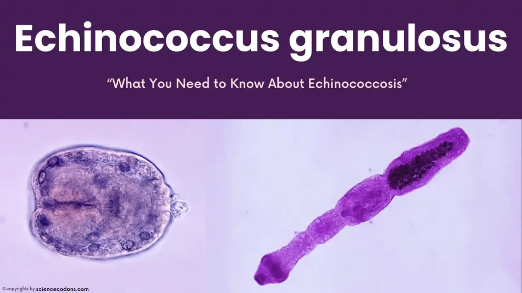
The worm is small, measuring 3-9 millimeters in length. Its scolex, or head, is spherical and armed with 4 suckers, a rostrum, and 2 rows of hooks. The worm has a short neck and a lifespan of 5-12 months. Echinococcus granulosus has 3 to 4 segments (one immature segment, one mature segment, and one fertile segment). The fertile segment forms about half the worm’s length and is critical to its identification. The worm has a single ovary with 2 lobes and 45-65 eggs. The genital opening is lateral and irregular, and the uterus has 12-15 branches. The eggs of Echinococcus granulosus are similar to other taenias.
The larval stage of the worm is a hydatid cyst, which appears as a single cavity and is spherical. It is known as a hydatid due to the fluid inside it. The cyst has a diameter of approximately 5 centimeters. It comprises different layers, including a laminated layer, a germinal layer inside, and primary heads (protoscolex) that bud out from this germinal layer. Also, broad capsules, a single-layer germinal layer, come out of the germinal layer, which contains protoscolexes.
Sometimes, in the mother cyst, there is a cyst exactly similar to the mother cyst, which is called the daughter cyst. It has both a germinal layer and primary heads (proto-scolexes) and is only located inside the mother cyst of Echinococcus granulosus. The primary heads have 2 states: either the head is inside (Invaginate), or the head is outside (Evaginate). Hydatid sand is a collection of primary heads and daughter cysts that settle at the bottom of the cyst and have a sandy appearance. The result of the host body’s immune response to the presence of the cyst is fibrosis. These fibrous layers are formed outside the laminated layer. The broad capsule is visible at the bottom, and the primary heads, which mostly have the invaginate state, are inside it.
Epidemiology
echinococcus granulosus is the causative agent of cystic echinococcosis, a zoonotic disease with a worldwide distribution. The epidemiology of E. granulosus is closely linked to domestic and wild canids, which act as definitive hosts, and livestock such as sheep, cattle, and goats, which are intermediate hosts.
Global Distribution: The parasite is endemic in many parts of the world, including Africa, Europe, Asia, the Middle East, Central and South America, and in rare cases, North America1. The prevalence of infection in these areas varies, influenced by factors such as animal husbandry practices, the presence of suitable definitive hosts, and human behaviors that facilitate transmission.
Life Cycle of Echinococcus granulosus
The life cycle of the tapeworm Echinococcus granulosus involves two hosts. The primary hosts are dogs and dog-like animals; the intermediate hosts are cattle, sheep, and herbivores. Humans can also be accidental intermediate hosts. Infected dogs, which are mostly stray dogs, excrete segments full of eggs. These segments disintegrate on grass and vegetables, releasing the eggs.
If herbivores consume this contaminated grass, or if humans accidentally eat contaminated vegetables, the embryo emerges from the egg. It reaches various organs, including the liver, lungs, and brain, through the bloodstream. Here, it forms a hydatid cyst within three weeks. If dogs or dog-like animals eat these infected organs of herbivores, adult worms form in the dog’s intestine for each primary head that is inside the cyst. The time to form adult worms is 6-9 weeks.
Pathogenicity and Symptoms
The pathogenesis of Echinococcus granulosus depends on the size, number, and location of the cyst, host sensitivity, growth pattern, etc. Most cysts form in the liver, as the blood from the intestines first goes to the portal vein and eventually goes to the liver. Therefore, 60-70 percent of hydatid cysts are in the liver, 20-40 percent are in the lungs, 4 percent are in the muscles, and 3 percent are in the spleen and brain.
- Liver cysts: Most are seen in the anterior right lobe of the liver. Their symptoms are abdominal pain, obstructive jaundice (due to obstruction of the bile ducts), and fever.
- Lung cysts: Symptoms include chest pain, chronic cough, shortness of breath, bloody sputum, and the presence of primary heads in the sputum.
- Bone cysts: These cysts are mostly reticulated—the cyst forms in the bone marrow. We usually do not have a limiting fibrous membrane; they have a small amount of fluid, and the primary heads in these cysts are few.
- Brain cysts: It causes symptoms such as Jacksonian epilepsy, increased intracranial pressure, and severe headaches.
In addition to the symptoms mentioned for hydatidosis caused by Echinococcus granulosus, another symptom is ascites or water belly due to the loss of body proteins and cyst growth. The size of the cysts varies and can be the size of a volleyball that divides the brain into two halves.
| Organ Involved | Symptoms |
|---|---|
| Liver | Abdominal pain, jaundice |
| Lungs | Chest pain, coughing up blood |
| General | Hives, anaphylaxis |
Diagnosis
the history of the disease, a history of contact with dogs, eating vegetables contaminated with dog feces, and clinical symptoms help in diagnosis.
Definitive diagnosis
This is possible by microscopic observation of protoscoles obtained from cyst fluid aspiration. Other symptoms are anemia and eosinophilia (20-25 percent). Imaging techniques such as radiology, CT scan, and MRI can be used to diagnose cysts.
Casoni test
They aspirate the cyst fluid of Echinococcus granulosus and inject some of it under the patient’s skin. After 24-48 hours, they check, and if there is redness, erythema, and swelling more than 5 millimeters, they say the person is positive. This test is not accurate and has a cross-reaction. Serological tests such as IHA and ELISA are also used, which also have a cross-reaction with cysticercosis Taenia solium and are inaccurate.
Treatment of Echinococcus
For the treatment of Echinococcus granulosus, the primary choice is surgery. The mortality rate for the disease is 1 to 4 percent. Along with surgery, Albendazole can be used at a dose of 12.5 milligrams per kilogram for three to six months. Praziquantel can be used at a dose of 120-210 milligrams per kilogram for six days before surgery. Flubendazole can be used at a dose of 50 milligrams per kilogram for 3-6 months.
Prevent Echinococcosis
It’s necessary to disrupt its transmission chain and life cycle to prevent infection with this parasite. The following measures can be taken to achieve this:
- Eliminate stray dogs: Stray dogs are often carriers of the parasite, so their elimination can help reduce the spread of the infection.
- Treat herd and pet dogs: Dogs that are part of a herd or kept as pets should be treated with medications such as Praziquantel and Arecoline to kill any parasites they may carry.
- Thoroughly wash vegetables and fruits: Proper washing of vegetables and fruits can remove any eggs or larvae of the parasite that may be present.
- Avoid feeding dogs with offal contaminated with hydatid cysts: This is a common practice in slaughterhouses, but it can spread the parasite.
- Vaccinate livestock: Livestock such as cows and sheep can be vaccinated to protect them from parasites.
- Fence off vegetable gardens and farms: This can prevent stray dogs, which may be carriers of the parasite, from entering these areas and contaminating the produce.
Conclusion
Echinococcus granulosus is a parasitic tapeworm with a complex lifecycle that impacts animals and humans. Understanding its transmission, lifecycle, and prevention strategies is crucial for managing and reducing the incidence of hydatid disease. Public health efforts, veterinary control measures, and community education are key components in combating this parasitic infection.
Reference:
- CDC
- https://doi.org/10.1136/bmj.e3866
- Nabarro LE, Amin Z, Chiodini PL: Current management of cystic echinococcosis: a survey of specialist practice. Clin Infect Dis. 60(5):721-8, 2015. doi: 10.1093/cid/ciu931.
- Horton RJ: Albendazole in treatment of human cystic echinococcosis: 12 years of experience. Acta Trop64(1-2):79-93, 1997. doi:10.1016/s0001-706x(96)00640-7
- Human Alveolar Echinococcosis after Fox Population Increase, Switzerland – PMC (nih.gov)
